
Machine Vision Fundus Camera Solutions
Capture more detailed, more consistent fundus images by incorporating JADAK’s leading machine vision technology into the fundus imaging process.
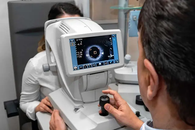
Enhance fundus imaging with JADAK’s machine vision technology, designed to aid ophthalmologists and optometrists in capturing more consistent, better detailed images of the interior surface of the eye. Our area scan machine vision cameras can be used in digital fundus imaging to support the diagnosis and monitoring of retinal conditions.
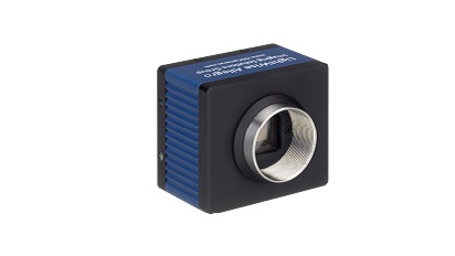
The Allegro USB machine vision camera series incorporates field programmable gate array (FPGA) technology to support real-time fundus imaging, engineered by one of the industry’s leading FPGA design teams. JADAK’s highly experienced engineers have developed more than 200 FPGA designs since 2000, transforming everything from surgical imaging to automated optical inspection.
Custom Fundus Imaging Solutions
Build a fundus camera that integrates easily with your existing system electronics to increase overall performance while reducing system costs. JADAK can source the best sensor for your fundus imaging needs from multiple vendors, and incorporate custom form factors including board level development and I/O.
Through close collaboration with your engineering team, the JADAK team integrates custom image processing, custom modes and other processing into the FPGA built for your fundus camera to reduce sensor noise while supporting image pre-processing.
Common Ophthalmic Imaging Challenges
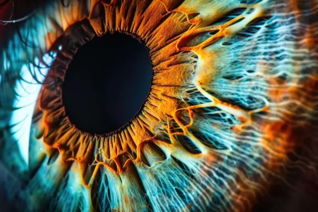
- Image Quality: It can be difficult to ensure consistency of image resolution and quality with some fundus imaging technology, which can impact diagnosis and monitoring.
- Small Field of View: If the field of view isn’t wide enough, this affects how detailed and comprehensive fundus imaging can be, potentially hindering diagnosis.
JADAK’s Fundus Camera Solutions
Key Features:
- In-built field programmable gate array (FPGA) for real-time fundus image processing
- Large image buffers and advanced processing capability
- LightWise Image Processing for superior image quality
- Custom image processing and manipulation options
- Advanced sensor correction
- Ideal for enhanced fundus visualization
- Lightweight and compact
Benefits of Machine Vision in Fundus Imaging
- Improved Image Consistency: Utilizing machine vision in an OCT fundus camera provides consistent image quality, as the technology reduces the variability caused by different imaging conditions.
- Wider Field of View: JADAK’s machine vision area scan cameras capture high-resolution, wide-field images in one shot. By covering more of the eye’s surface without the requirement for multiple images, as fundus imaging is more efficient than other ophthalmic imaging methods.
- Reliable Diagnosis and Monitoring: The key benefit of machine vision fundus imaging is that the improved image resolution it enables supports the reliable diagnosis and monitoring of eye conditions, subsequently benefiting patient outcomes.
JADAK Healthcare Imaging Solutions
Our machine vision surgical imaging solutions include OCT imaging, fundus imaging, microscopy imaging, and surgical navigation systems. Capture higher quality images to benefit patient diagnoses. Contact us today to discuss our healthcare engineering solutions.
Frequently Asked Questions (FAQs)
A fundus camera provides enhanced image quality and consistency compared to other ophthalmic imaging solutions. Machine vision area scan cameras can capture a wider field of view, showing a greater level of detail at a higher resolution. This type of digital fundus imaging can therefore support more accurate and reliable patient diagnoses and monitoring.
A fundus camera will capture highly detailed images of the rear of the eye, which includes the retina, optic disc, fovea, macula and posterior pole. Fundus imaging can show details such as blood vessel abnormalities, helping ophthalmologists to identify conditions faster and more accurately.
An ophthalmic fundus camera is primarily used in applications such as retinal photography and fluorescein angiography.
During retinal photography, a fundus camera is used to capture images of the interior surface of the eye to detect and manage conditions including diabetic retinopathy, glaucoma, and age-related macular degeneration.
In fluorescein angiography, fundus imaging can highlight certain retinal landmarks, aiding diagnosis.
JADAK Customized Engineering
With our engineer-to-engineer collaboration approach, our team becomes an extension of yours. It starts by gaining a deeper understanding of your application. Then, we’ll help develop and implement a solution that’s a precise fit, supporting you throughout the product lifecycle.