
Machine Vision OCT Imaging Solutions
Enhance the resolution and quality of retinal images using industry-leading machine vision technology to enhance the OCT imaging process.
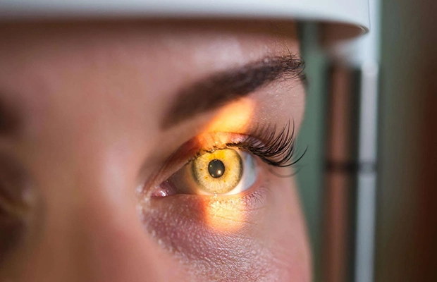
Capture high-resolution retinal images faster during OCT imaging by using machine vision line scan cameras to take highly detailed cross-sectional images of eye tissue. A line camera scans the retina one pixel line at a time to rapidly create high-resolution images, supporting more accurate patient diagnoses via OCT retinal imaging.
JADAK’s engineers will work with you to build a machine vision OCT camera tailored to your precise needs, featuring FPGA (field programmable gate array) technology to enable real-time image processing. We’re proud to have one of the imaging industry’s top FPGA design teams, who have successfully developed more than 200 FPGA-based products since 2000. Get in touch to discover how JADAK’s leading technology can transform your OCT imaging system technology.
OCT Imaging Solutions
Create an Optical Coherence Tomography (OCT) imaging system tailored to your precise needs; engineer-to-engineer collaboration means all of JADAK’s healthcare engineering solutions are fully customizable. View each of the retina’s distinctive layers more clearly when using a machine vision-powered OCT camera, which can capture high-resolution images during retinal imaging and optic nerve analysis.
Integrate system electronics and custom technology into the OCT camera’s FPGA, and customize form factors including board level, cooled sensors and custom I/O to develop a unique OCT imaging machine to support enhanced monitoring and more accurate diagnosis of eye conditions.
OCT Imaging Challenges
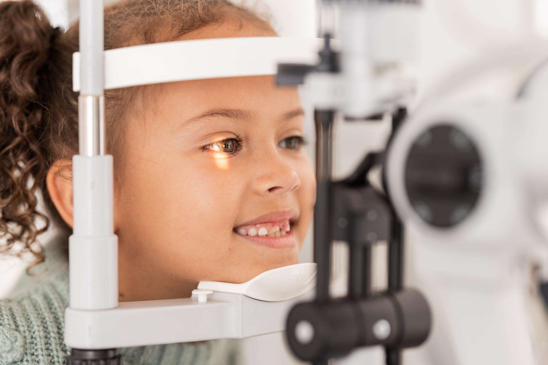
- Image Resolution: Low-resolution, poor-quality images can hinder the timely, accurate diagnosis of retinal conditions, impacting patient outcomes.
- Processing Speed: Slow image processing speeds can lead to greater periods of discomfort for patients, increasing the likelihood of motion artifacts, affecting overall image quality.
Machine Vision OCT Camera Solutions
Warning: Undefined variable $custom_block_styles in /home/customer/www/jadaktech.boldfocus.com/public_html/wp-content/themes/Jadak2022/blocks/accented-content-block/accented-content-block.php on line 60
Key Features:
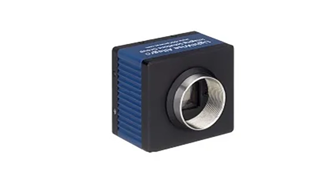
- Capture fast-moving images in high resolution
- Real-time image processing supported by in-built field programmable gate array (FPGA) technology
- Reduce motion artifacts associated with patient movement by speeding up the OCT imaging process
- Fully customizable and scalable for ease of integration with existing equipment, reducing cost and enhancing performance
Benefits of Machine Vision in OCT Medical Imaging
- Higher Resolution Images: Machine vision line scan cameras capture high-resolution, highly detailed images during OCT retinal imaging, supporting the more accurate diagnosis and monitoring of eye conditions, benefiting patient outcomes.
- Faster Image Processing: Line scan cameras capture images at a significantly faster speed than traditional cameras, making OCT imaging a much faster process. This in turn minimizes the risks associated with motion artifacts caused by patient movement.
Whitepaper: Powerful Vision Systems Using Modular FPGA-Based Smart Cameras
JADAK Healthcare Imaging Systems
JADAK’s machine vision and RFID technology solutions can be used in a range of healthcare applications, including surgical imaging, endoscopy, microscopy imaging, and in-vitro diagnostics. Contact our team to discuss how our industry-leading technology can support your OCT medical imaging requirements.
Frequently Asked Questions (FAQs)
Using machine vision technology as part of an OCT imaging system enhances the quality and resolution of cross-sectional retinal images. This supports more accurate, timely diagnosis, meaning patients can access the right treatment faster, benefiting overall outcomes.
An OCT imaging system featuring machine vision can be used in applications including retinal imaging and optic nerve analysis.
During retinal scans, OCT imaging is used to capture highly detailed cross-sectional retinal images to support the diagnosis and monitoring of conditions such as glaucoma, macular degeneration, and diabetic retinopathy.
In optic nerve analysis, an OCT camera can evaluate the optic nerve fibers, helping to detect changes caused by glaucoma and other eye conditions.
The main benefit of Optical Coherence Tomography (OCT) imaging over Magnetic Resonance Imaging (MRI) scanning is that an OCT camera provides significantly higher resolution images showing a greater level of detail, supporting more accurate diagnosis. Another key difference of OCT medical imaging vs MRI scanning is that the former relies on light to capture images, while the latter uses radiofrequency technology.
JADAK Customized Engineering
With our engineer-to-engineer collaboration approach, our team becomes an extension of yours. It starts by gaining a deeper understanding of your application. Then, we’ll help develop and implement a solution that’s a precise fit, supporting you throughout the product lifecycle.